
From diagnosing disease under the microscope to developing the next generation of cancer therapies, artificial intelligence is reshaping the way healthcare professionals and researchers approach complex problems. As AI continues to evolve, its greatest potential lies not in replacing human expertise but in enhancing it. This article explores how AI is transforming cancer pathology and biomedical research through current advances and my own experiences in both a hospital pathology department and a university research laboratory.
Artificial Intelligence in Healthcare
Artificial intelligence has become deeply embedded in modern society, with applications spanning countless industries and even our personal lives. From predicting financial trends for major businesses to providing grammatical feedback on student writing, AI has an enormous capacity to automate and optimize both complex and routine tasks. One field where this optimization has become increasingly valuable is healthcare.
Currently, artificial intelligence lacks the clinical judgment, intuition, and experience that physicians develop through years of education and practice. Rather than replacing healthcare professionals, AI serves as a powerful decision-support tool by automating repetitive tasks, identifying patterns in large datasets, and streamlining patient care.
Artificial intelligence has proven especially promising in precision medicine, particularly in medical imaging and diagnostics. AI algorithms can analyze X-rays, MRIs, and CT scans to identify early signs of diseases such as cancer, stroke, and hidden bone fractures. One study by two UK universities found that an AI model accurately diagnosed strokes in more than 2,000 patients and could even estimate when the stroke had occurred—an essential factor in determining appropriate treatment.
Beyond medical imaging, AI systems can continuously monitor patient data to identify individuals at high risk for serious medical events before symptoms appear. They can also detect subtle biological signatures that may predict future disease. Researchers have already demonstrated AI’s ability to identify early indicators of conditions including Alzheimer’s disease, chronic obstructive pulmonary disease (COPD), and kidney disease.
AI in Cancer Pathology
AI has transformed many aspects of healthcare, with one of its most promising applications being cancer pathology. There is currently a global shortage of pathologists, particularly in low- and middle-income countries. At the same time, traditional pathology relies on the careful microscopic examination of thousands of tissue samples—a process that is time-consuming, labor-intensive, and occasionally subject to variability between observers.
To improve both efficiency and consistency, AI systems can objectively analyze digital pathology slides and assist pathologists in making standardized diagnoses. For example, AI models have been shown to assign Gleason scores more consistently than human pathologists. The Gleason score is a grading system used to determine the severity of prostate cancer.
In addition to improving consistency, AI has demonstrated impressive diagnostic accuracy. Studies have shown that AI can detect cancer in particularly challenging cases, such as identifying lymph node metastases in breast cancer or recognizing subtle early-stage lung cancer lesions. Furthermore, AI systems can evaluate patients more comprehensively by integrating multiple forms of clinical data, including genomic sequences, medical histories, and radiologic images.
Traditional pathology services can also be difficult to access in rural or underserved communities, delaying diagnoses and potentially life-saving treatments. Similar to telemedicine, AI-powered digital pathology platforms enable pathologists to collaborate remotely, increasing access to specialized expertise while improving both the speed and accuracy of diagnosis.
My Pathology Experience
Last summer, I had the opportunity to intern in a pathology department at Rochester General Hospital, where I experienced the diagnostic process firsthand. I examined a variety of histological slides—from prostate cancer to stomach polyps—and observed how pathologists analyze tissue architecture to arrive at a diagnosis.
Each slide required several minutes to examine and even longer to formally document. Although the pathologist I shadowed was remarkably experienced and could often recognize disease almost instantly, reviewing slide after slide remained a time-intensive and highly repetitive process that demanded sustained focus.
Occasionally, I was asked to diagnose cases myself using textbooks and online references. Even with labeled images and step-by-step diagnostic guides, distinguishing healthy tissue from malignant tissue proved surprisingly difficult. Characterizing subtle differences in cellular architecture was even more challenging.
Another component of my internship involved reviewing patient medical histories for evidence of thyroid cancer and using genomic sequencing software to identify associated genetic mutations. Ultimately, our analysis found strong genotype-phenotype correlations in thyroid cytology, suggesting that gene-level information may help predict histologic diagnoses following surgical resection.
Given these findings, artificial intelligence has the potential to recognize patterns between genetic signatures and cytologic outcomes. It could also help personalize treatment by predicting treatment responses, identifying patients who may benefit from targeted therapies, and matching individuals to mutation-specific clinical trials. These capabilities could significantly streamline the therapeutic decision-making process.
AI in Cancer Research
In addition to my pathology internship, I conduct cancer research in Dr. Julie Pollock’s biochemistry laboratory at the University of Richmond. I maintain three cancer cell lines as shown below: H23 lung (top), T-47D breast (middle), and U-87 MG glioblastoma (bottom). While the glioblastoma cells display a distinct elongated morphology, the lung and breast cancer cells are nearly indistinguishable to the naked eye.
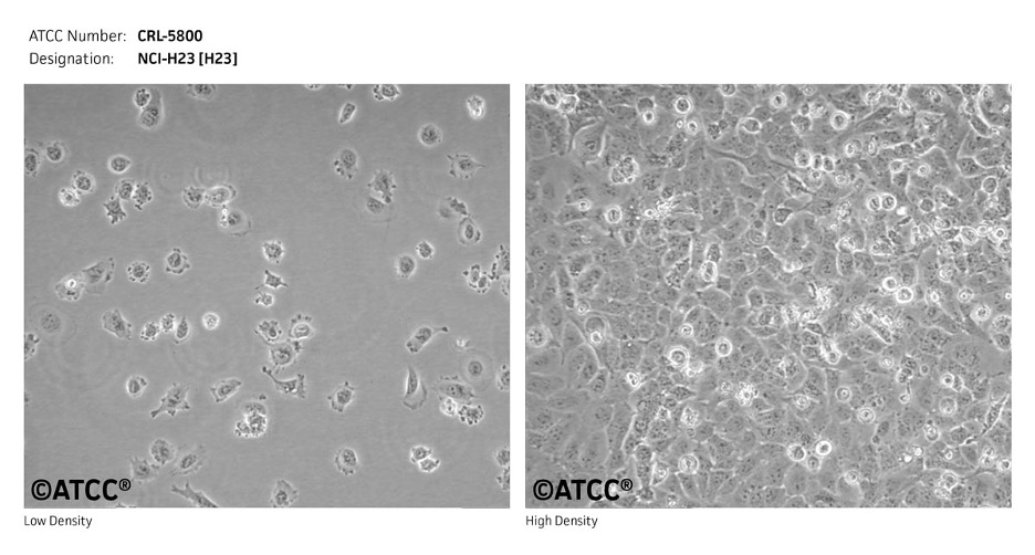
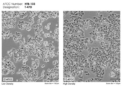
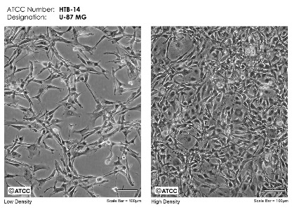 Images from ATCC.
Determining the optimal time to passage cells based on confluency can also be challenging. AI-assisted image analysis could more accurately distinguish between cell lines, quantify cell confluency, and determine the ideal time for passaging. These improvements would help ensure consistent cell culture practices and increase the reliability of downstream experiments.
My research focuses on photoactivated chemotherapy, an emerging approach that uses light to activate an initially inert prodrug directly within a tumor. I conduct cell viability assays using various ruthenium compounds and ligands to determine which combinations of compound concentration and light wavelength most effectively kill cancer cells.
Artificial intelligence also has the potential to streamline this research by predicting cellular responses before experiments are performed. AI models could identify the most promising compound-ligand combinations, optimize drug concentrations and light exposure parameters, analyze large cell viability datasets, and detect subtle patterns that researchers might otherwise overlook.
By reducing the number of experiments required, AI could accelerate the development of safer, more selective photoactivated therapies while allowing researchers to focus their efforts on the most promising candidates. Ultimately, these advances have the potential to reduce both the time and cost associated with developing novel anticancer treatments.
Looking Ahead
Artificial intelligence is transforming the way cancer is diagnosed, studied, and treated, but its greatest value lies in complementing—not replacing—the expertise of physicians, pathologists, and researchers. Through my experiences in both a hospital pathology department and a cancer research laboratory, I have seen firsthand the time, precision, and collaboration required to advance cancer care.
As AI continues to evolve, it has the potential to reduce routine workloads, uncover insights that might otherwise go unnoticed, and help guide more informed clinical and research decisions. By combining the strengths of human expertise with artificial intelligence, the future of cancer care can become not only more efficient, but also more precise and personalized.
Written by Piper Turri, a Biology and Neuroscience student at the University of Richmond.
Recent Comments